Diagnostic Ultrasound in Manhattan and NYC

Diagnostic ultrasound may help evaluate certain joint, tendon, ligament, muscle, nerve, foot, abdominal wall, hernia-related, and pelvic-region pain patterns.

Learn how we use diagnostic ultrasound to evaluate muscles, tendons, nerves, and joints, helping identify the source of pain and guide personalized treatment.

What Is Diagnostic Ultrasound?

Diagnostic ultrasound is an imaging tool that uses sound waves to view certain soft tissue structures in real time. In pain medicine, it may help evaluate joints, tendons, ligaments, muscles, nerves, superficial soft tissue changes, foot pain, abdominal wall pain, hernia-related pain, and selected pelvic-region pain patterns.

At Manhattan Pain Medicine, diagnostic ultrasound is used as part of a diagnosis-first evaluation, not as a standalone answer for every pain condition. It can provide useful information when symptoms require targeted, dynamic, anatomy-specific, or procedure-planning assessment, but it does not replace clinical evaluation or other imaging when needed.

Specialist-Guided Ultrasound Evaluation

MPM specialists use diagnostic ultrasound to help clarify selected pain generators when the clinical picture supports it. This may include tendon pain, ligament injury, joint irritation, chronic sprain, nerve entrapment, foot pain, shoulder pain, hip pain, knee pain, abdominal wall pain, hernia-related pain, or superficial pelvic-region pain patterns.

Ultrasound findings are interpreted in context with symptoms, exam findings, prior imaging, medical history, functional limitations, and treatment goals. The goal is not simply to perform another test, but to determine whether ultrasound can help refine diagnosis, guide care decisions, or support safer and more accurate procedure planning.

Real-Time Imaging for Pain Evaluation

Diagnostic ultrasound can be especially useful when pain changes with movement, pressure, position, or specific activity. Unlike static imaging, ultrasound allows the clinician to view certain structures in real time and, in some cases, assess how tendons, ligaments, muscles, joints, nerves, or soft tissue structures behave during motion.

This can be helpful for patients with persistent pain after prior X-rays, MRI, CT, physical therapy, injections, or specialist visits, especially when the pain source remains unclear. Ultrasound may support evaluation of tendinopathy, chronic sprain, shoulder impingement, frozen shoulder, foot pain, Morton’s neuroma, tarsal tunnel syndrome, peripheral nerve entrapment, slipping rib syndrome, abdominal wall pain, hernia-related pain, or selected pelvic-region symptoms. At MPM, ultrasound is used within a broader diagnostic framework. Some conditions still require MRI, CT, X-ray, EMG, lab work, vascular imaging, rheumatology care, neurology care, pelvic specialty evaluation, or surgical assessment.

Request An Appointment

Evaluation Process

How MPM Uses Diagnostic Ultrasound

MPM uses diagnostic ultrasound when real-time imaging may help clarify the pain source or support treatment planning.
  • 1

    Review the Pain Pattern

    The process begins with a careful review of pain location, triggers, movement limits, prior injuries, prior imaging, medical history, exam findings, and treatment response. MPM considers whether symptoms suggest a joint, tendon, ligament, muscle, nerve, soft tissue, foot, abdominal wall, hernia-related, or pelvic-region pain generator.
  • 2

    Determine Ultrasound Fit

    Diagnostic ultrasound is not the right test for every condition. MPM determines whether ultrasound can reasonably evaluate the suspected structure or whether MRI, CT, X-ray, EMG, lab work, vascular imaging, or specialist evaluation may be more appropriate.
  • 3

    Perform Targeted Imaging

    When appropriate, ultrasound is used to examine selected anatomy in real time. The evaluation may include static and dynamic views, comparison with movement, assessment of superficial soft tissue structures, or identification of anatomy relevant to treatment planning.
  • 4

    Connect Findings to Care

    MPM interprets ultrasound findings in clinical context. Results may help guide next steps, such as rehabilitation, medication management, additional imaging, specialist referral, ultrasound-guided injections, or a different diagnostic pathway if ultrasound does not explain the symptoms.

Diagnostic Ultrasound Across MPM’s Pain Expertise

Diagnostic ultrasound may support several MPM Zones of Expertise, including Complex Chronic Pain, Hypermobility, Autoimmune and Inflammatory pain, Musculoskeletal issues, and Pelvic Pain. In musculoskeletal care, ultrasound may help evaluate selected tendon, ligament, muscle, joint, and soft tissue pain patterns. In hypermobility-related pain, it may help assess certain dynamic or soft tissue contributors when clinically relevant. In autoimmune or inflammatory pain, ultrasound may help identify selected joint or tendon findings that require broader clinical interpretation.

For pelvic-region, abdominal wall, hernia-related, foot, ankle, shoulder, hip, knee, or nerve entrapment symptoms, ultrasound may help clarify whether a superficial structural or soft tissue pain generator is involved. MPM uses ultrasound selectively, as part of a coordinated diagnostic plan rather than as a replacement for deeper medical evaluation.

Learn More

What to Expect During Diagnostic Ultrasound

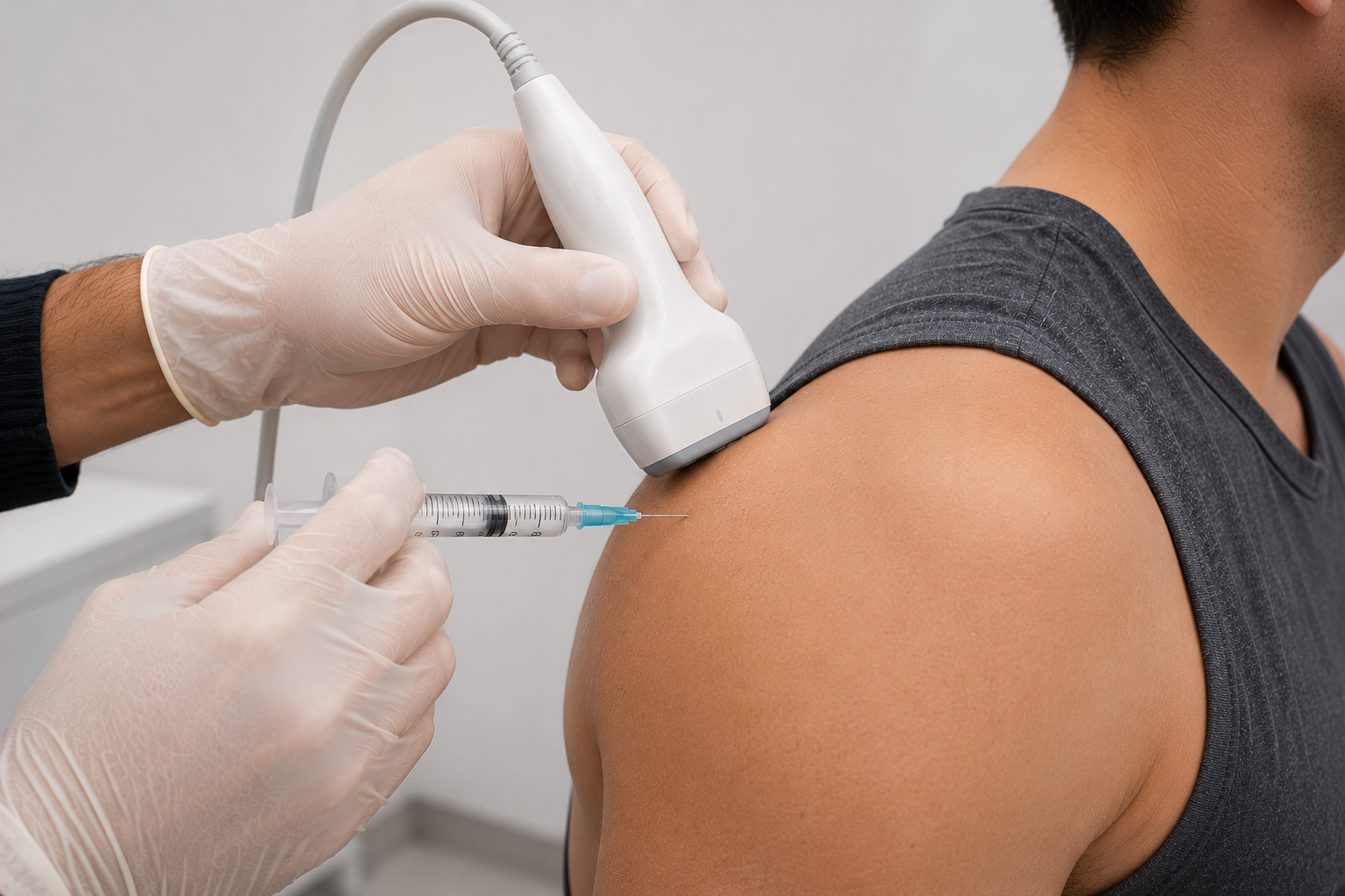
During a diagnostic ultrasound exam, a clinician applies gel to the skin and uses a handheld probe to view selected structures. The exam may focus on a joint, tendon, ligament, muscle, nerve, soft tissue area, foot or ankle region, abdominal wall, hernia-related area, or superficial pelvic-region concern depending on the symptoms. In some cases, the patient may be asked to move, change position, or reproduce a symptom while the area is viewed in real time.

Diagnostic ultrasound does not use radiation and is usually well tolerated. Some patients may feel mild pressure from the probe, especially over a painful area. After the exam, MPM reviews findings in the context of symptoms and clinical goals. If ultrasound does not explain the pain, additional imaging or specialist evaluation may still be needed.

Request An Appointment

Related Conditions

Conditions Evaluated With Diagnostic Ultrasound

Diagnostic ultrasound may help evaluate selected pain conditions depending on the suspected structure, symptoms, exam findings, and clinical context.
PATIENT STORIES

Real Patients. Real Progress.

Hear from patients who came to Manhattan Pain Medicine looking for answers, clarity, and a more thoughtful path forward.
  • Madaline. M

    5 star review

    I cannot say enough good things about Manhattan Pain Medicine. From the moment I walked in, I felt truly cared for and taken seriously. Dr. Nino Mikaberidze, Director of Rheumatology, is exceptional — knowledgeable, compassionate, and incredibly thorough. She takes the time to listen, explains every...

    Google

  • Haleigh Y.

    5 star review

    The best care I've received as a chronic pain patient. I traveled from out of state to visit Manhattan Pain Medicine because I could not find knowledgeable doctors in my hometown for my hypermobility, and Ehlers-Danlos syndrome symptoms and comorbidities. Let me tell you, it was worth every travel e...

    Google

  • Damien P.

    5 star review

    I was injured in a bicycle accident over 8 years ago. I’ve suffered throughout the years since my accident with nothing seeming to work. PT, Yoga, lifestyle changes, NOTHNG! I finally made an appointment with Dr Kane and I’m feeling an improvement in my life. I no longer stay up all night in pain, a...

    Google

  • JP S.

    5 star review

    One of the best experiences, if not the best, I have ever had at the doctor’s office. From the front desk to the nurses to the PA assisting the doctor this was a great experience. Dr.Nino actually listened to me and came up with a game plan. Would recommend this practice to as many people as I could...

    Google

  • Richard B.

    5 star review

    I have very unique & severe nerve pain from CRPS which has no known specific cure. Dr. Siefferman has a huge tool box, far greater than the other 23 Drs. I have seen. He saved me from jumping off a bridge and continues to help me with both treatments and advice. Dr. S. is in a different league!

    Google

  • Clare F.

    5 star review

    After two years of searching and feeling lost and let down, I finally found real help at Manhattan Pain Medicine. With Dr. Siefferman, Adam Rosenberg, Dr. Mikaberidze, and the whole team, I feel heard, supported and understood. Every symptom is being considered and I’m finally getting answers with m...

    Google

  • Jonathan C.

    5 star review

    Why is Dr Dr. Tayyaba Ahmed one of the top pevic Floor specialists - 1- She wants to know a full history of what led the patient to get to this point. 2 - She listens intensley to the patient 3- She does not rush you 4- Extremely skilled at giving pelvic floor injections 5- I thank my luck stars she...

    Google

  • Jodi K.

    5 star review

    The staff was friendly and helpful. Dr. Siefferman was thoughtful, listened, asked questions and proposed simple safe ways to come to further conclusions regarding my pain and discomfort. The office staff including Dr. Siefferman is responsive even after the initial in-person visit. Very pleased.

    Google

  • Sabrina S.

    5 star review

    Dr. Siefferman is a brilliant provider. While my pain is still a work in progress, Dr Siefferman always gives me options and explains his reasoning behind things. He listens to my feedback and concerns. I never feel rushed; and am grateful he works to meet me where I am. I highly recommend him.

    Google

  • Lorraine B.

    5 star review

    What a great find! They listen to me, I listen to them and the diagnosis is made and a plan developed to develop an approach to treat all of my chronic pain. I wish I could have discovered this type of treatment sooner. This doctor and group give me hope.

    Google

FAQs About Diagnostic Ultrasound

Related Ultrasound-Guided Treatments

Related treatments may be considered depending on the ultrasound findings, suspected pain generator, diagnosis, and treatment goals.

Understanding Insurance Before You Begin

Before care begins, our team helps patients understand the practical side of treatment, including insurance verification, out-of-network benefits, cost-share estimates, self-pay options, and billing questions. We believe patients should have as much clarity as possible before moving forward.

Insurance & Billing

Pain Care That Starts With Understanding

When pain is complex, the first step should not be another generic treatment. Manhattan Pain Medicine looks deeper to understand what is driving the pain, why it has persisted, and what path forward makes sense for you.

Request An Appointment

Insights & Research

Explore MPM Insights and Research, including patient-friendly articles, clinical perspectives, and research from our team to help explain how we understand complex pain, evaluate care options, and guide the path to treatment
In Depth

Diagnostic Ultrasound for Pain Evaluation

Diagnostic ultrasound may help clarify selected pain generators when used within a diagnosis-first clinical evaluation.

Diagnostic Ultrasound

Diagnostic ultrasound is a real-time imaging tool that uses sound waves to view selected structures beneath the skin. In pain medicine, it may help evaluate certain joints, tendons, ligaments, muscles, nerves, soft tissue structures, foot and ankle problems, abdominal wall pain, hernia-related pain, and superficial pelvic-region pain patterns.

At Manhattan Pain Medicine (MPM), diagnostic ultrasound NYC care is part of discovery-led, diagnosis-first pain evaluation. It is not used as a standalone answer for every pain condition. Instead, it is used when the clinical question fits what ultrasound can evaluate and when the findings may help guide treatment decisions.

How Diagnostic Ultrasound Helps Evaluate Pain

Many patients seek diagnostic ultrasound after persistent pain has not improved with physical therapy, medication, injections, prior imaging, or specialist care. Some have had normal or inconclusive X-rays, MRI, CT, or nerve studies but still have symptoms that seem tied to movement, pressure, position, or a specific soft tissue structure.
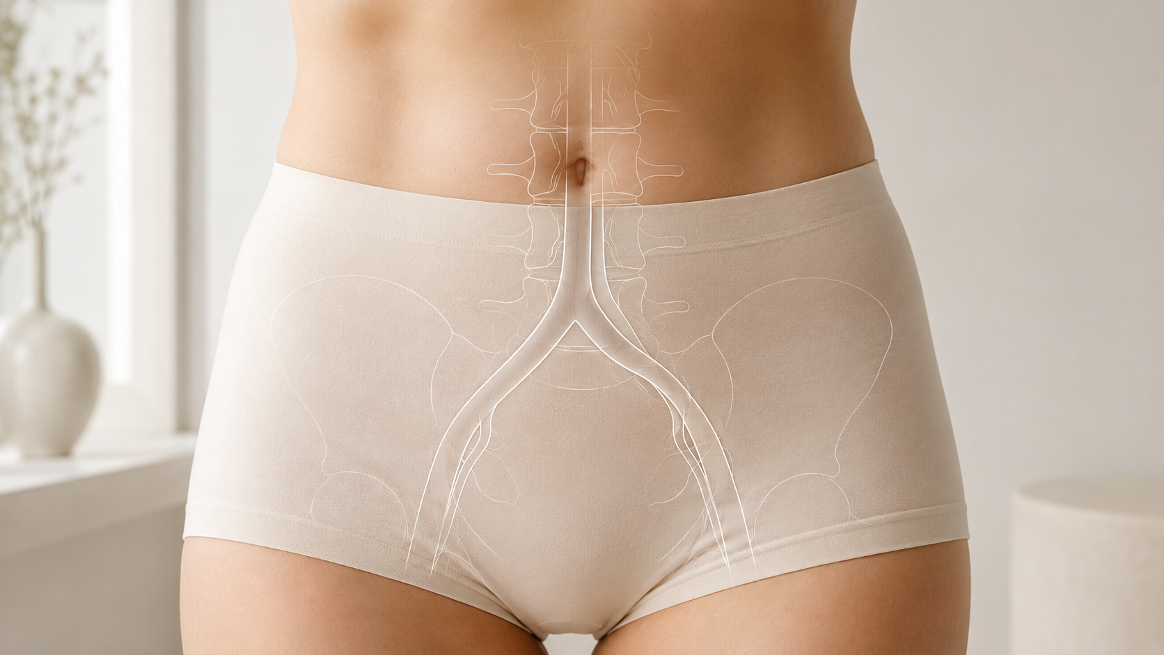

Diagnostic ultrasound may help assess selected pain generators in real time. This can include tendons, ligaments, muscles, joints, superficial nerves, soft tissue masses, fluid collections, bursae, or areas of irritation. It may also help compare anatomy during movement, which can be useful when pain changes with activity.


Diagnostic Ultrasound Versus MRI, CT, and X-Ray

Ultrasound is different from MRI, CT, and X-ray. X-ray is useful for many bone and joint alignment questions. CT can provide detailed bone and structural imaging. MRI can evaluate many deep structures, joints, soft tissues, nerves, discs, and internal anatomy. Ultrasound is often strongest for real-time, high-resolution evaluation of accessible soft tissue structures and dynamic movement.

Because each test answers different questions, ultrasound should be chosen based on the suspected diagnosis. For example, ultrasound may help evaluate tendinopathy, chronic sprain, shoulder impingement, Morton’s neuroma, tarsal tunnel syndrome, or superficial nerve entrapment in selected cases. MRI or CT may be needed for deeper joint, spine, pelvic, abdominal, or neurological concerns.

Musculoskeletal Ultrasound for Joints, Tendons, and Ligaments

Musculoskeletal ultrasound may help evaluate joint, tendon, ligament, and muscle pain in selected areas. This may include shoulder impingement, frozen shoulder, hip or knee pain, meniscal or labral concerns where appropriate, patellofemoral knee pain, scapular dyskinesia, tendinopathy, chronic sprain, TFCC injury, slipping rib syndrome, or soft tissue pain after injury.

Ultrasound can also help evaluate certain inflammatory findings, such as joint fluid or synovial changes, when clinically relevant. However, autoimmune and inflammatory conditions such as rheumatoid arthritis, lupus, Sjogren’s, vasculitis, and undifferentiated connective tissue disease require broader medical evaluation. Ultrasound may support the picture, but it does not replace rheumatology care, lab work, or systemic assessment.

Ultrasound for Nerve, Foot, Abdominal Wall, and Pelvic-Region Pain

Diagnostic ultrasound may help evaluate selected nerve-related pain patterns, including peripheral nerve entrapment, peroneal nerve entrapment, cubital tunnel syndrome, Morton’s neuroma, tarsal tunnel syndrome, or cluneal neuralgia. It may show certain nerve changes or surrounding soft tissue factors, but EMG, nerve conduction studies, MRI, or neurological evaluation may still be needed.

For foot pain, ultrasound may help assess soft tissue, tendon, ligament, nerve, or neuroma-related concerns. For abdominal wall pain, it may help evaluate selected hernia-related pain patterns, sports hernia concerns, abdominal wall structures, or superficial nerve entrapment when clinically appropriate. For pelvic-region pain, ultrasound may help answer targeted musculoskeletal or superficial soft tissue questions, but it does not replace evaluation for pelvic floor dysfunction, pudendal neuralgia, endometriosis, vascular compression syndromes, urologic conditions, gynecologic conditions, or gastrointestinal causes.

Dynamic Ultrasound and Injection Planning

One of ultrasound’s strengths is dynamic evaluation. The clinician may observe certain structures while the patient moves, changes position, or reproduces a symptom. This can be useful when pain occurs only during activity or when static imaging does not fully explain the complaint.

Ultrasound may also support procedure planning. Before ultrasound-guided injections, it can help identify anatomy, nearby nerves, blood vessels, soft tissue structures, or target areas. During selected procedures, ultrasound guidance may help improve needle placement and procedural safety. MPM uses this selectively when it fits the diagnosis and treatment goal.

When Ultrasound May Not Be Enough

Diagnostic ultrasound is operator-dependent and should always be interpreted in clinical context. It cannot diagnose every pain condition. It is not definitive for many deep spine conditions, intracranial pressure disorders, Chiari malformation, CSF outflow obstruction, jugular stenosis, tethered cord, complex pelvic organ disease, autoimmune disease, or vascular compression syndromes. Additional imaging, lab work, vascular studies, EMG, specialist evaluation, or emergency care may be needed.

Patients should seek urgent evaluation for sudden severe headache, new neurological deficits, chest pain, shortness of breath, severe abdominal or pelvic pain, bowel or bladder dysfunction, saddle anesthesia, fever, unexplained weight loss, major trauma, severe swelling, suspected infection, loss of pulses, rapidly worsening weakness, or rapidly worsening symptoms.

For selected patients, diagnostic ultrasound can be a valuable part of pain evaluation. MPM’s role is to determine when ultrasound can answer the right clinical question and how the findings should guide the next step in coordinated care.