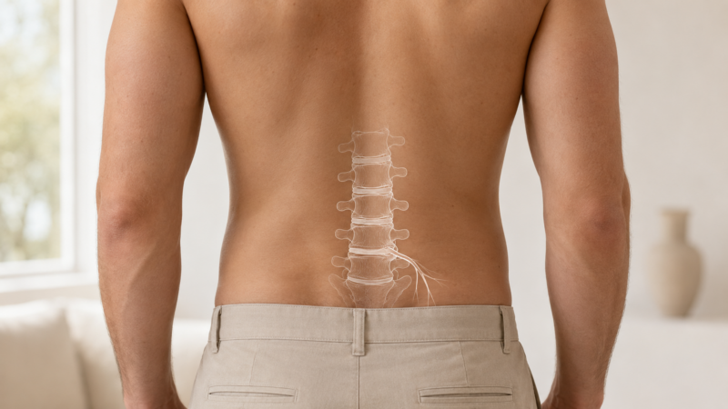

Many patients arrive with an MRI report showing a “mild disc bulge,” yet the pain is severe. Others have large disc herniations on imaging and very little discomfort.
This is why an MRI alone cannot explain the full pain story.
A herniated disc and a pinched nerve are related, but they are not the same thing. Leg pain, groin pain, hip pain, numbness, tingling, or burning may come from mechanical pressure, chemical irritation, nerve tension, or a combination of factors.
The “Pinched Nerve” Problem
The term “pinched nerve” can make it sound as if the nerve is simply being squeezed. Sometimes that happens, but many disc-related symptoms are driven by inflammation.
A spinal disc has a tougher outer ring and a softer inner material. When the outer ring stretches, sprains, or tears, inflammatory disc material can irritate nearby nerve roots. The nerve may become painful even when the compression looks mild on imaging.
This is one reason a small disc finding can create significant symptoms, while a larger finding may not bother another patient at all.
Disc Pain vs. Nerve Pain
Disc-related pain can show up in different ways.
Radicular pain occurs when a nerve root is irritated. This may cause sharp, electric, burning, or zapping pain that travels down the leg, often called sciatica.
Discogenic pain comes from the disc itself. When the outer ring of the disc is injured, pain may be felt in the low back, hips, groin, pelvis, or legs. This can be harder to localize and may be mistaken for a hip, pelvic, or muscle problem.
Why the MRI May Miss the Driver
A standard MRI is taken while the patient is lying still. It does not always show what happens when the spine is standing, bending, twisting, or carrying weight.
A disc that looks mild while lying down may irritate a nerve during movement or sitting. Scar tissue can also develop as a disc heals, sometimes tethering the nerve and limiting its ability to glide. This nerve tension may not be obvious on imaging.
In some patients, there may also be a double crush pattern. A nerve can be irritated in the spine and then placed under tension again downstream near the piriformis, pelvis, or hip.
What a Proper Workup Requires
A proper workup should not treat the MRI report alone. It should connect the imaging to the symptoms, physical exam, movement patterns, and nerve tension testing.
In some cases, targeted epidural treatment may help calm inflammation and improve nerve mobility. Rehabilitation then becomes important to address the mechanics that placed stress on the disc in the first place.
Our Approach at MPM
At Manhattan Pain Medicine, we evaluate disc pain and nerve pain through a diagnosis-first lens, helping patients understand whether symptoms are coming from disc inflammation, nerve irritation, scar tissue, instability, or a layered spine and pelvis pattern.