Meniscal Tear
A meniscal tear is an injury to one of the menisci, the C-shaped cartilage structures that sit between the thighbone and shinbone. Each knee has two menisci. They help cushion the joint, distribute load, and support knee stability. When a meniscus is torn, patients may feel pain, swelling, stiffness, clicking, catching, locking, giving way, or difficulty fully straightening the knee.
A meniscal tear may happen suddenly during a twisting or pivoting movement, especially when the foot is planted and the knee rotates. It may also occur during sports, deep squatting, kneeling, or trauma. In other patients, the tear develops gradually as the meniscus weakens with age, arthritis, or degeneration.
Traumatic vs. Degenerative Meniscal Tears
A traumatic meniscal tear often happens during a specific injury. The patient may remember a twist, pop, or sudden pain, followed by swelling, stiffness, or difficulty continuing activity. This is common in sports and activity-related knee injuries.
A degenerative meniscal tear may develop more gradually. Patients may not remember a single injury. Symptoms may appear with stairs, squatting, walking, twisting, or prolonged activity. Degenerative tears often occur in knees that also have arthritis or cartilage wear. People with knee arthritis may be more prone to meniscal tears.
This distinction matters because treatment may differ. A young athlete with a traumatic tear and mechanical symptoms may need a different care pathway than a patient with degenerative meniscal findings and knee arthritis.
Why Imaging Does Not Always Tell the Full Story
MRI can be helpful for identifying meniscal tears and related knee pathology. However, the presence of a meniscal tear on imaging does not always prove it is the main pain source. Some tears are highly symptomatic. Others may be incidental or part of a broader degenerative knee pattern.
MPM evaluates whether the imaging matches the symptoms. The exam considers pain location, swelling, range of motion, mechanical catching or locking, ligament stability, strength, gait, arthritis signs, patellofemoral pain, tendon involvement, hypermobility, and referred pain from the hip or spine. The goal is not to treat the MRI report. The goal is to treat the patient’s actual pain generator.
Meniscal Tear vs. Other Causes of Knee Pain
Knee pain can come from many sources. A patient with clicking or pain may have a meniscal tear, but similar symptoms can also come from arthritis, cartilage injury, ligament strain, patellofemoral pain, tendon irritation, bursitis, nerve irritation, referred hip pain, referred spine pain, or inflammatory arthritis.
True locking, where the knee cannot fully straighten because of a mechanical block, is different from occasional clicking or discomfort. True locking may require orthopedic evaluation, especially when it follows injury or is associated with significant swelling or loss of motion.
Hypermobility and Knee Instability
Hypermobility can make meniscal and knee symptoms more complex. Patients with EDS or hypermobility spectrum disorder may have increased joint laxity, altered movement control, and greater reliance on muscles for stability. This can place extra stress on ligaments, tendons, cartilage, and the meniscus.
In these cases, care may need to address stability, proprioception, load management, and movement patterns, not only pain. A patient with hypermobility-related knee pain may need a different plan than a patient with an isolated traumatic meniscal tear.
How MPM Evaluates Meniscal Tear Pain
MPM begins with a detailed history and physical examination. This includes how symptoms started, whether there was a twisting injury, where the pain is located, whether the knee swells, whether it catches or locks, whether the patient can fully straighten the knee, and what activities are limited.
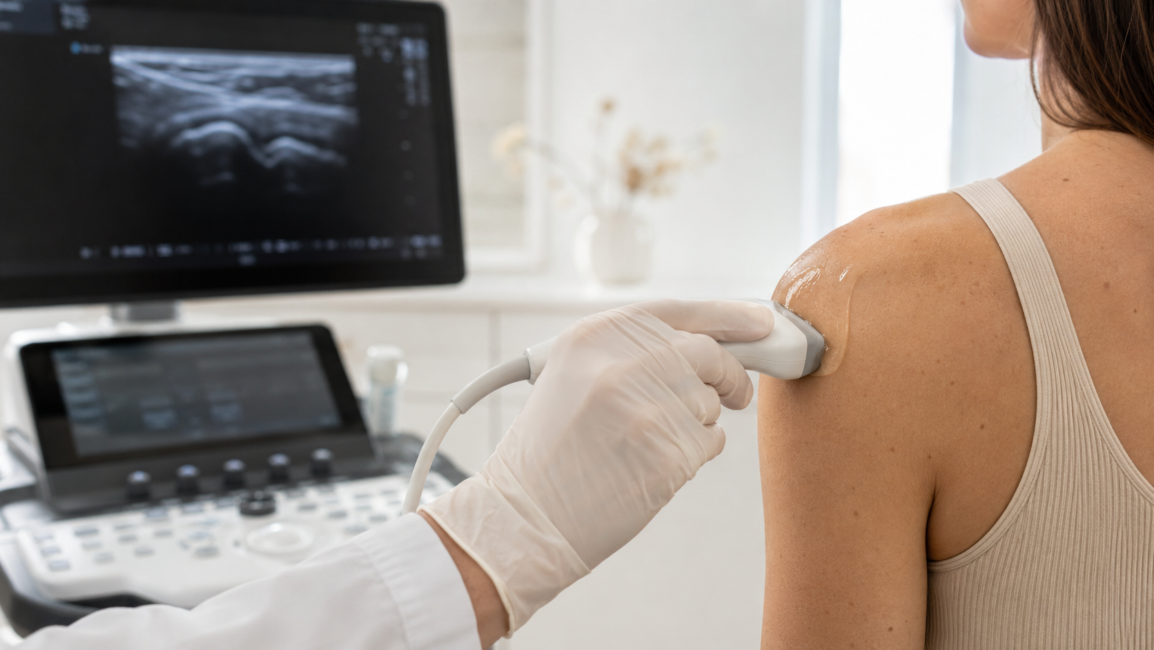
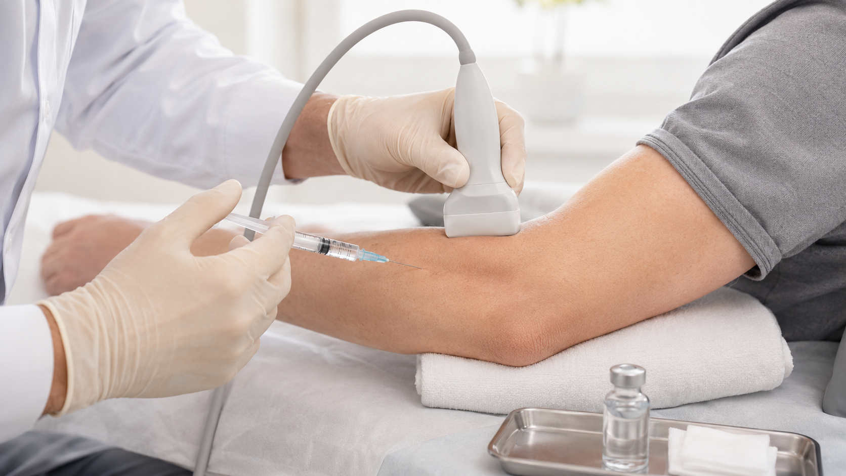
The exam may evaluate joint line tenderness, range of motion, swelling, ligament stability, patellar tracking, strength, gait, and functional movements such as squatting or stepping. Imaging is reviewed in context. MRI may be useful for internal meniscal detail. Diagnostic ultrasound may help assess selected findings such as effusion, meniscal extrusion, or surrounding soft tissue and may guide injections when appropriate.
Treatment Options for Meniscal Tears
Treatment depends on the tear type, symptoms, mechanical features, arthritis burden, age, activity goals, and knee stability. Conservative care may include rest, activity modification, avoiding twisting or pivoting, ice, compression, elevation, bracing, medication management, and physical therapy. Treatment often begins with avoiding aggravating activities and using supportive measures such as ice when appropriate.
Physical therapy may focus on restoring motion, reducing swelling, improving strength, improving control, and gradually returning to activity. For degenerative tears, the plan may also need to address arthritis and long-term load management.
Image-Guided Injections and Regenerative Options
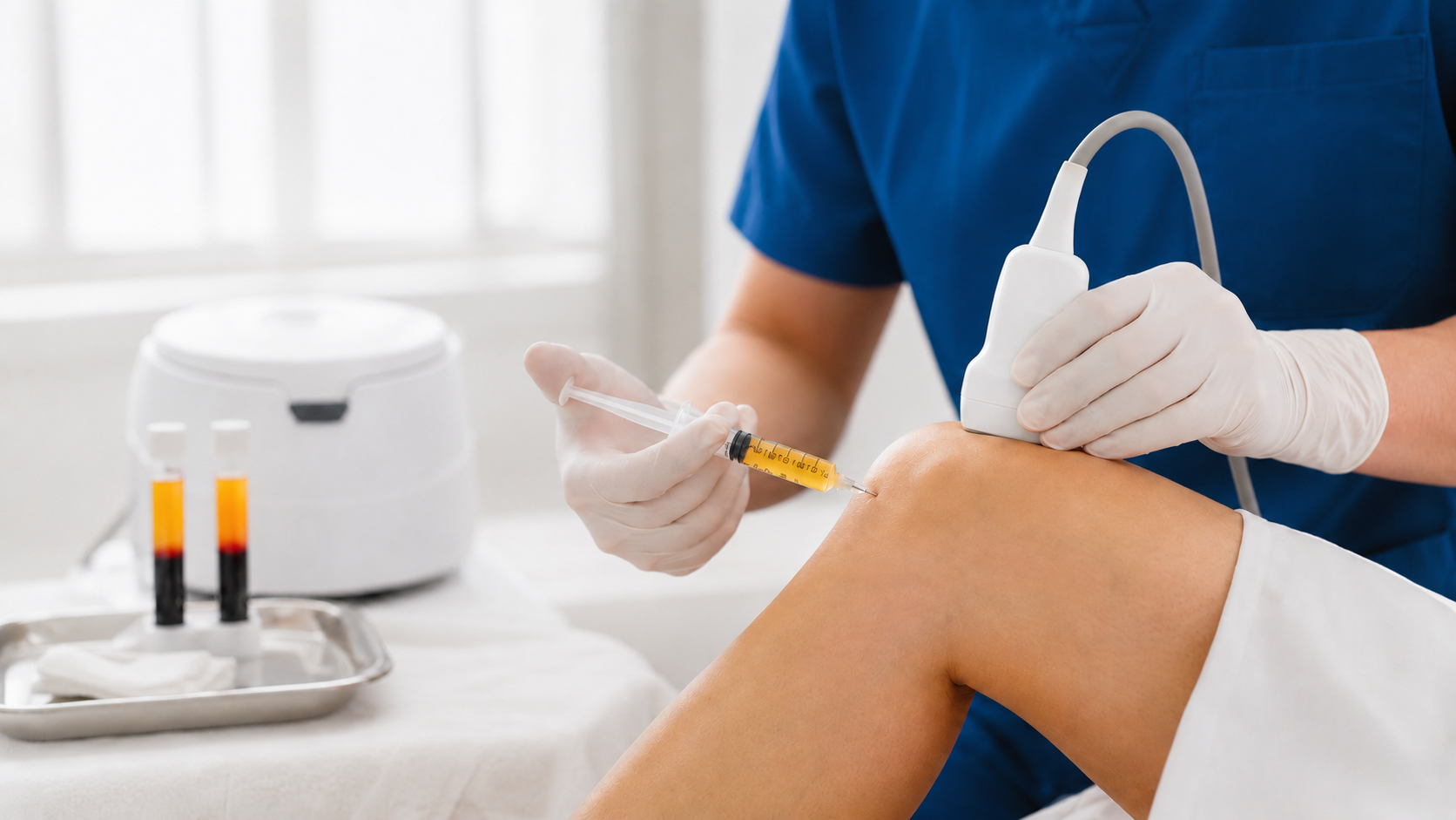
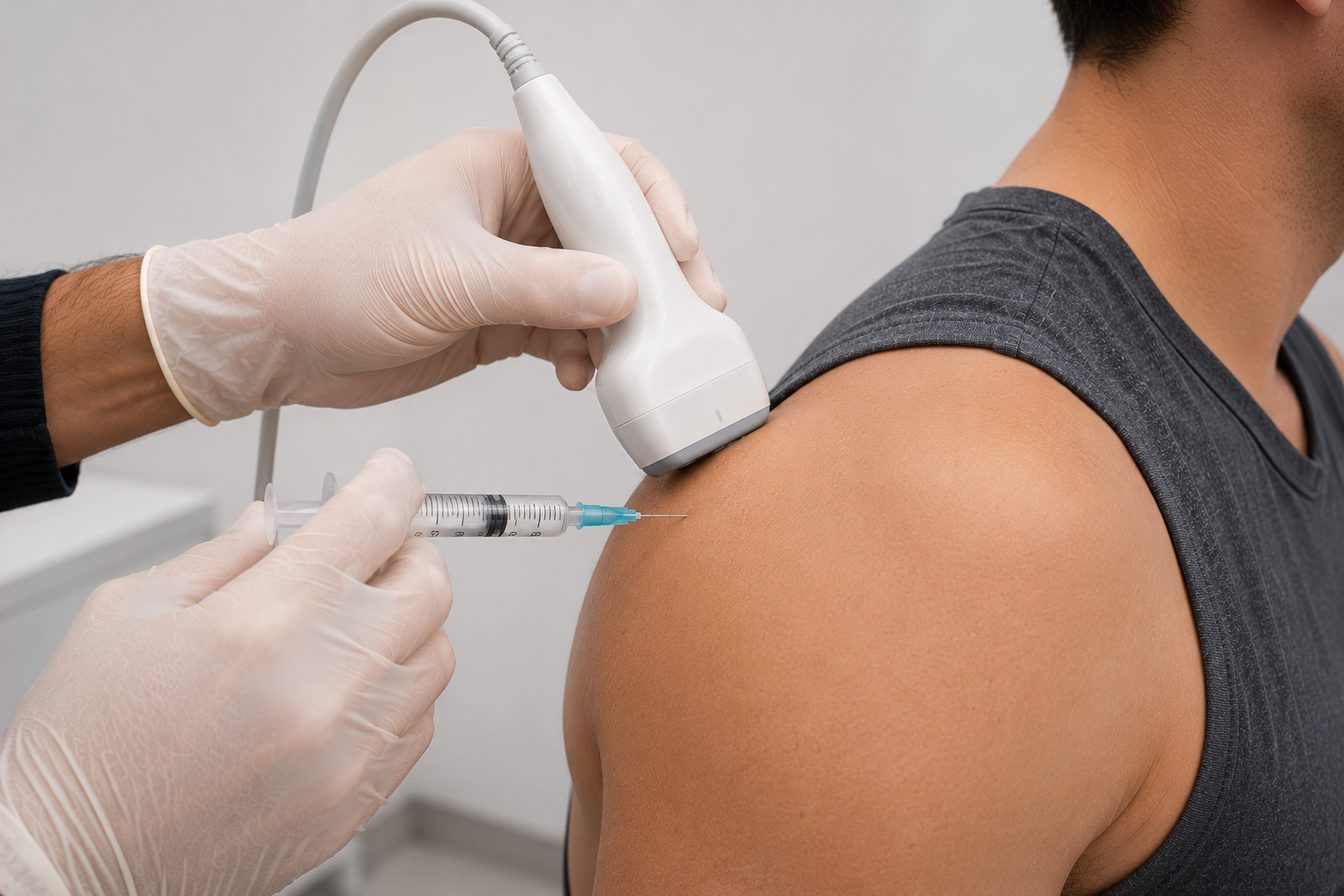
In selected patients, ultrasound-guided or image-guided injections may be considered for pain control, inflammation, or diagnostic clarification. Steroid injections may reduce inflammation and symptoms in selected cases but do not repair the tear itself. AAOS notes that physical therapy and corticosteroid injections may relieve symptoms but do not address the structural injury. (OrthoInfo)
Some patients ask about PRP, BMAC, prolotherapy, or regenerative medicine. These may be discussed only after reviewing the tear pattern, arthritis burden, anatomy, symptoms, goals, risks, and evidence. They should not be presented as guaranteed meniscus repair, cartilage restoration, arthritis reversal, or a substitute for surgery when surgery is medically indicated.
When Surgery May Be Needed
Orthopedic referral may be appropriate when there is true locking, inability to fully straighten the knee, major traumatic tear, suspected root tear, persistent mechanical symptoms, severe functional limitation, or failure of appropriate nonsurgical care. Surgery may also be needed when structural damage requires repair or when symptoms remain significant despite a careful treatment plan.
MPM does not frame surgery as failure. For selected patients, orthopedic evaluation and arthroscopy may be the appropriate next step.
When Knee Pain Needs Urgent Evaluation
Knee pain should be evaluated when symptoms persist, worsen, limit walking or activity, cause true locking, catching, instability, swelling, inability to fully straighten the knee, or follow trauma. Urgent evaluation is important for inability to bear weight, major swelling after injury, visible deformity, fever, redness, calf swelling, loss of pulses, new weakness or numbness, or rapidly worsening pain.
How MPM Approaches Meniscal Tear Care
MPM approaches meniscal tears through a diagnosis-first, coordinated model. The evaluation considers meniscal injury, arthritis, ligament instability, cartilage injury, tendon pain, hypermobility, EDS, referred hip or spine pain, peripheral nerve irritation, and chronic pain sensitization.
For patients looking for meniscus tear treatment in Manhattan, MPM offers a careful pain medicine perspective that can work alongside physical therapy, orthopedics, sports medicine, and rehabilitation. The goal is to determine whether the meniscal tear is the true pain generator, clarify whether nonsurgical care is appropriate, and coordinate referral when structural treatment may be needed.