Intracranial Hypertension
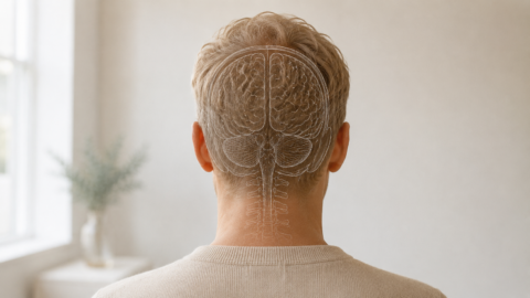
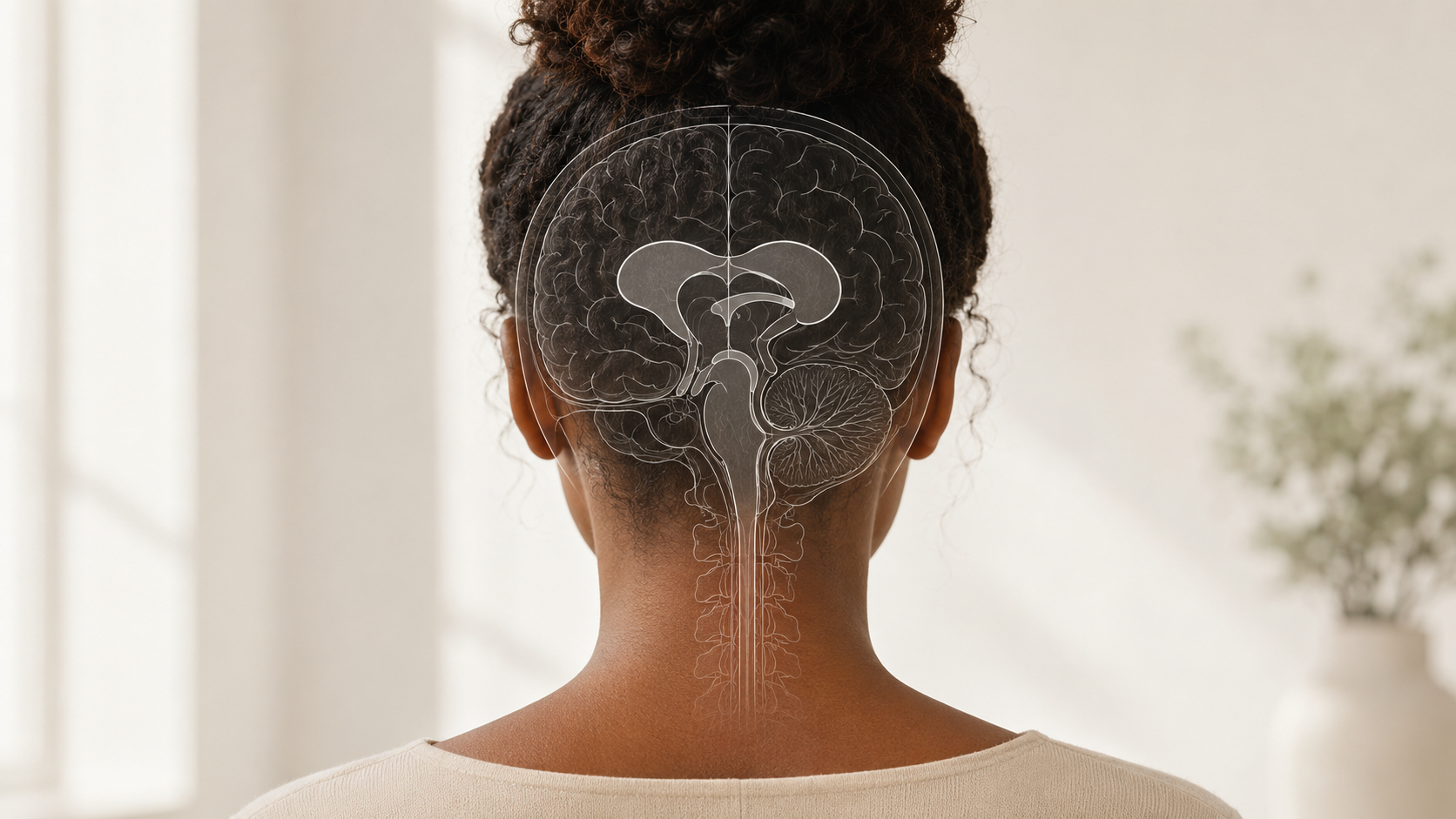
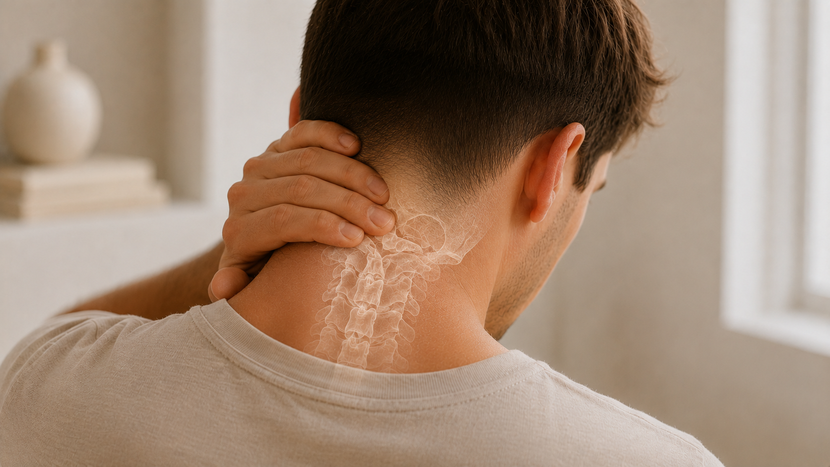
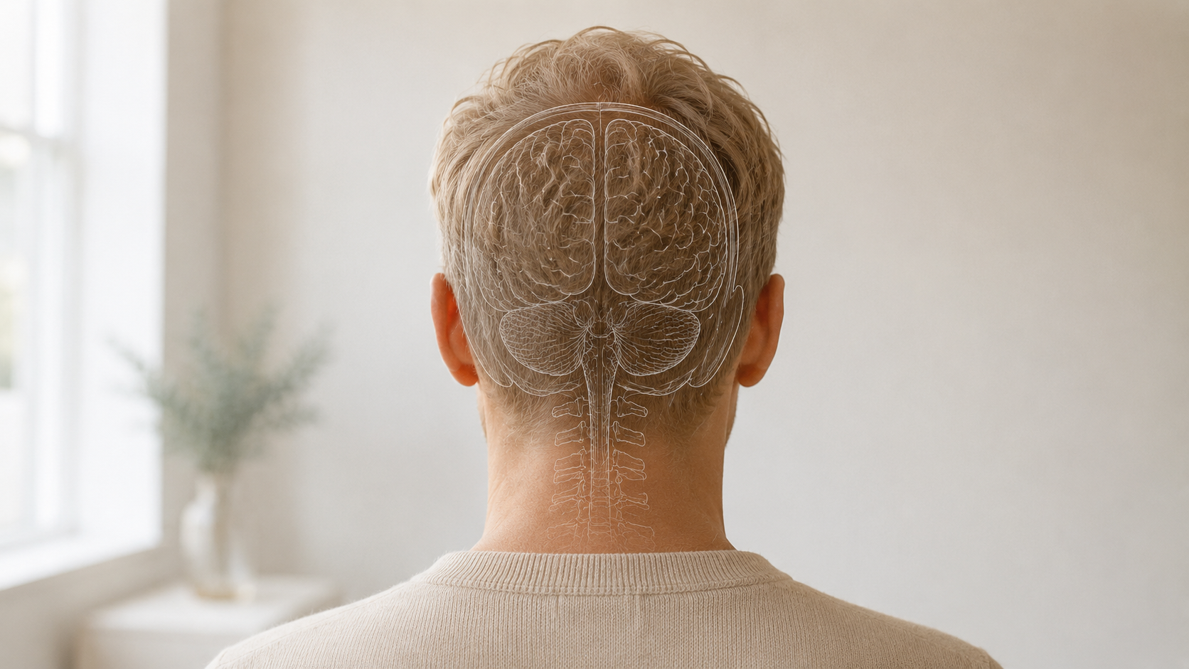
Intracranial hypertension means increased pressure inside the skull. This pressure may involve cerebrospinal fluid, brain blood flow, optic nerve structures, and the surrounding nervous system. When pressure is elevated, patients may experience headaches, nausea, pulsatile tinnitus, visual symptoms, dizziness, brain fog, and neck pain.
Idiopathic intracranial hypertension, or IIH, is a form of intracranial hypertension where pressure is elevated without a clear tumor or mass as the cause. It is also called pseudotumor cerebri. The word “idiopathic” means the cause is not clearly identified. Secondary intracranial hypertension refers to elevated pressure related to another cause, such as medication effects, venous outflow problems, endocrine factors, inflammatory conditions, or other medical issues.
Because intracranial hypertension can affect the optic nerves, suspected symptoms should be evaluated carefully and promptly, especially when vision changes are present.
Common Symptoms of Intracranial Hypertension
Symptoms may include pressure-like headache, nausea, vomiting, pulsatile tinnitus, blurry vision, double vision, brief episodes of vision dimming or loss, dizziness, neck pain, balance changes, light sensitivity, brain fog, and fatigue. Some patients report that headaches worsen when lying down, bending, coughing, straining, or changing position.
However, these symptoms are not specific to intracranial hypertension. Chronic migraine, vestibular migraine, Chiari malformation, CSF outflow obstruction, CSF leak, craniocervical instability, cervical spine disorders, POTS, EDS, HSD, post-COVID pain, inflammatory disease, medication effects, and chronic pain sensitization may create overlapping symptoms.
This is why MPM approaches suspected intracranial hypertension through diagnosis-first evaluation rather than assuming the cause based on symptoms alone.
Intracranial Hypertension and Vision Changes
Vision protection is one of the most important priorities in intracranial hypertension care. Elevated pressure can cause swelling of the optic nerves, called papilledema. Papilledema may lead to blurry vision, visual field loss, transient vision loss, double vision, or progressive visual symptoms.
A patient may have vision risk even if they can still read clearly or pass a simple vision check. Visual field testing, optic nerve examination, and neuro-ophthalmology evaluation may be important when papilledema or pressure-related vision changes are suspected.
MPM does not replace neuro-ophthalmology or emergency evaluation for vision-threatening symptoms. When progressive visual change or papilledema is suspected, timely referral or urgent evaluation is appropriate.
Intracranial Hypertension vs Migraine, Chiari, CCI, and CSF Disorders
Intracranial hypertension can be difficult to distinguish from other headache and neurologic conditions because symptoms may overlap.
Migraine can cause severe headache, nausea, light sensitivity, sound sensitivity, dizziness, visual symptoms, and functional impairment. Intracranial hypertension headaches may resemble migraine or tension-type headache, so headache quality alone is not enough to confirm the diagnosis.
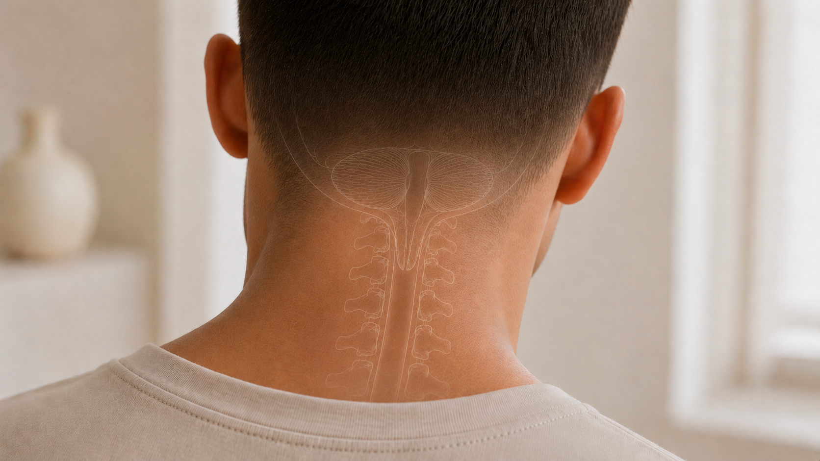
Chiari malformation can cause headache, neck pain, dizziness, balance symptoms, and CSF flow concerns in selected patients. Some Chiari symptoms may resemble intracranial hypertension, and some patients may require neurology or neurosurgery evaluation to clarify whether the Chiari finding is clinically meaningful.
Craniocervical instability can overlap with headache, neck pain, dizziness, visual symptoms, brain fog, and autonomic symptoms, especially in patients with EDS or HSD. CCI is different from intracranial hypertension, but both may be part of a broader evaluation in selected patients.
CSF outflow obstruction refers to impaired movement or drainage of cerebrospinal fluid. Intracranial hypertension refers to elevated pressure inside the skull. These conditions can overlap in some cases, but they are not the same.
How MPM Evaluates Suspected Intracranial Hypertension-Related Pain
MPM begins by reviewing the patient’s full symptom history, including headache pattern, pressure symptoms, positional changes, cough or strain sensitivity, pulsatile tinnitus, vision symptoms, nausea, dizziness, neck pain, brain fog, autonomic symptoms, prior imaging, eye findings, prior diagnoses, and treatment response.
The evaluation may include headache classification, musculoskeletal assessment, upper cervical pain mapping, trigger point and myofascial assessment, hypermobility-aware review, autonomic symptom screening, medication history, and review of red flags that require urgent or specialist evaluation.
MPM also reviews available imaging and specialist workups when provided. This may include MRI, MRV, eye exam findings, visual field testing, lumbar puncture history, opening pressure results, neuro-ophthalmology notes, neurology notes, or neurosurgical recommendations.
When intracranial hypertension is suspected or not yet fully evaluated, MPM may coordinate with neuro-ophthalmology, neurology, neurosurgery, neuroradiology, primary care, cardiology, genetics, rheumatology, ENT, vestibular therapy, or other clinicians.
Treatment Options for Intracranial Hypertension-Related Headache and Pain
Treatment depends on the diagnosis, severity, visual findings, and suspected cause. For idiopathic intracranial hypertension, specialist-directed care may include weight management when medically appropriate, medication to reduce CSF production, monitoring of vision, and treatment of headache morbidity.
In selected cases, lumbar puncture, shunting, venous sinus stenting, optic nerve sheath fenestration, or other neurosurgical or procedural care may be considered by the appropriate specialists. These are not routine pain medicine treatments and should be guided by neuro-ophthalmology, neurology, neurosurgery, or vascular and imaging findings.
MPM’s role is focused on pain and function. When patients have overlapping chronic migraine, upper cervical pain, myofascial pain, autonomic dysfunction, hypermobility-related mechanics, or chronic pain sensitization, MPM may support non-surgical pain management. This may include medication coordination, headache-directed care, acupuncture, Feldenkrais, biofeedback, pain psychology, trigger point injections, and selected image-guided procedures for clearly identified pain generators.
Pain psychology and biofeedback may be helpful when chronic symptoms create fear, vigilance, sleep disruption, medical trauma, stress physiology, or difficulty reengaging with care. These tools do not mean symptoms are imagined. They can support nervous system regulation, coping, pacing, and function while medical evaluation continues.
Image-Guided and Interventional Options
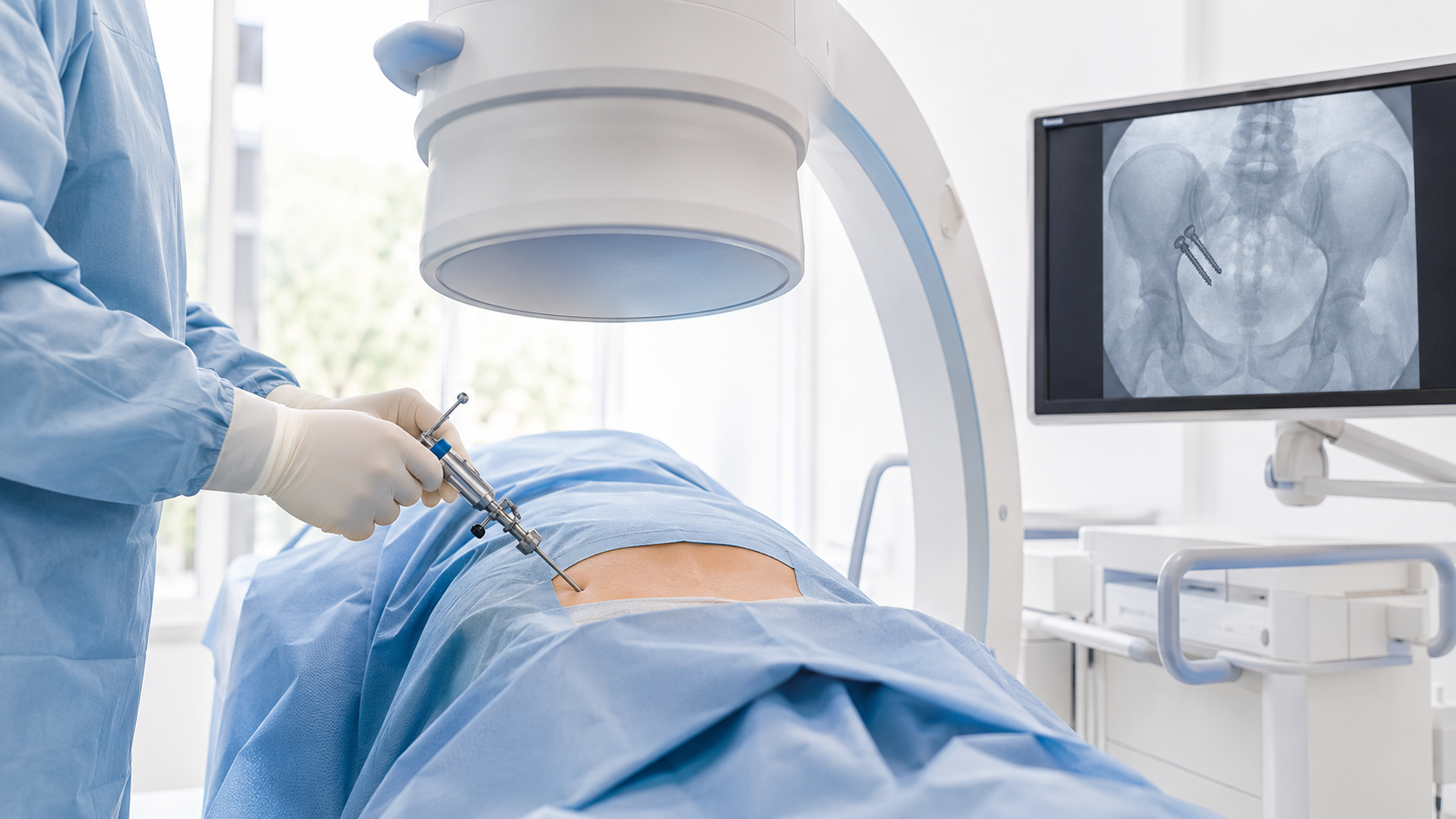
Some patients with suspected intracranial hypertension also have separate musculoskeletal or upper cervical pain generators. In those cases, targeted procedures may be considered only when the diagnosis supports them.
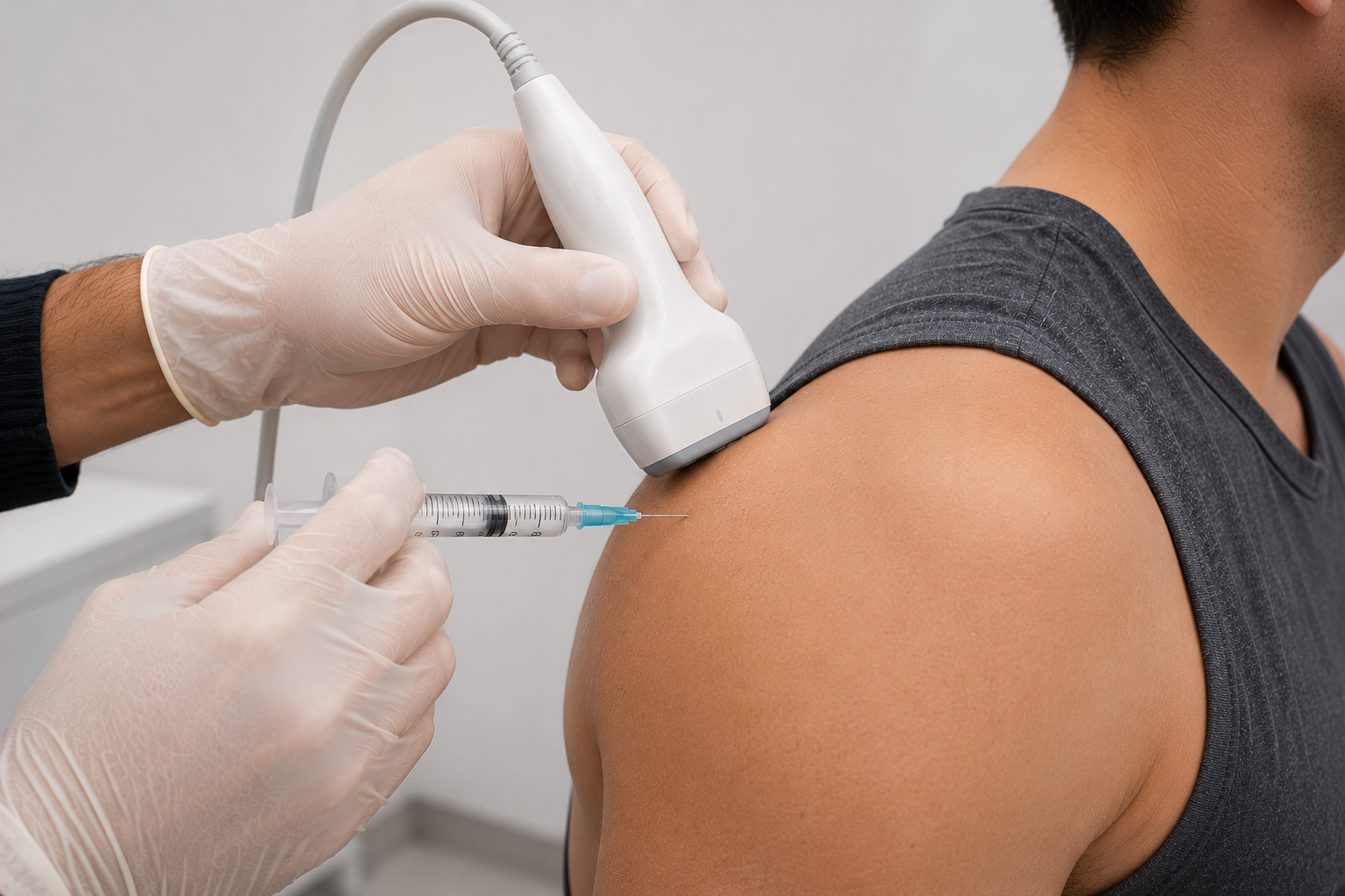
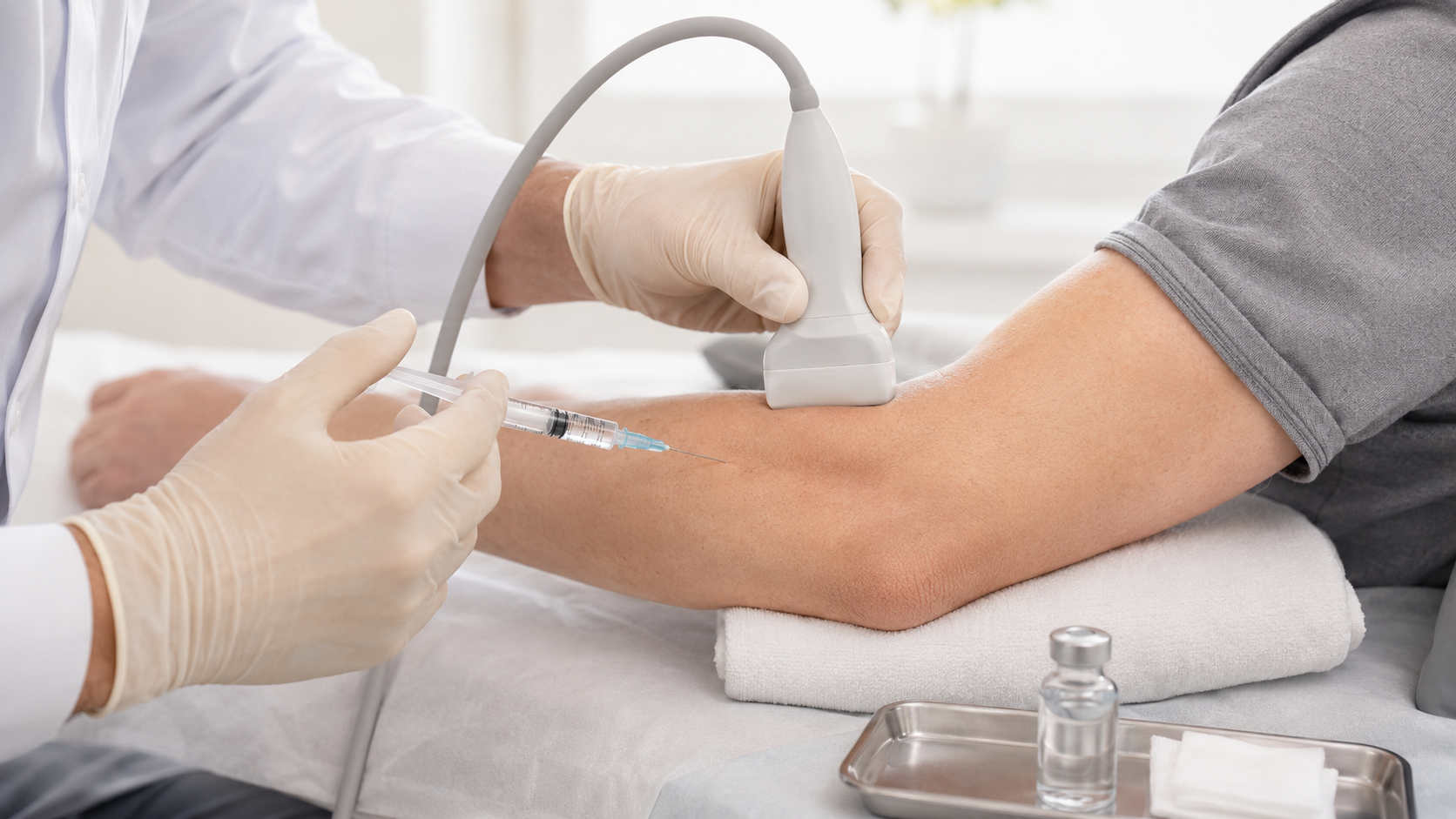
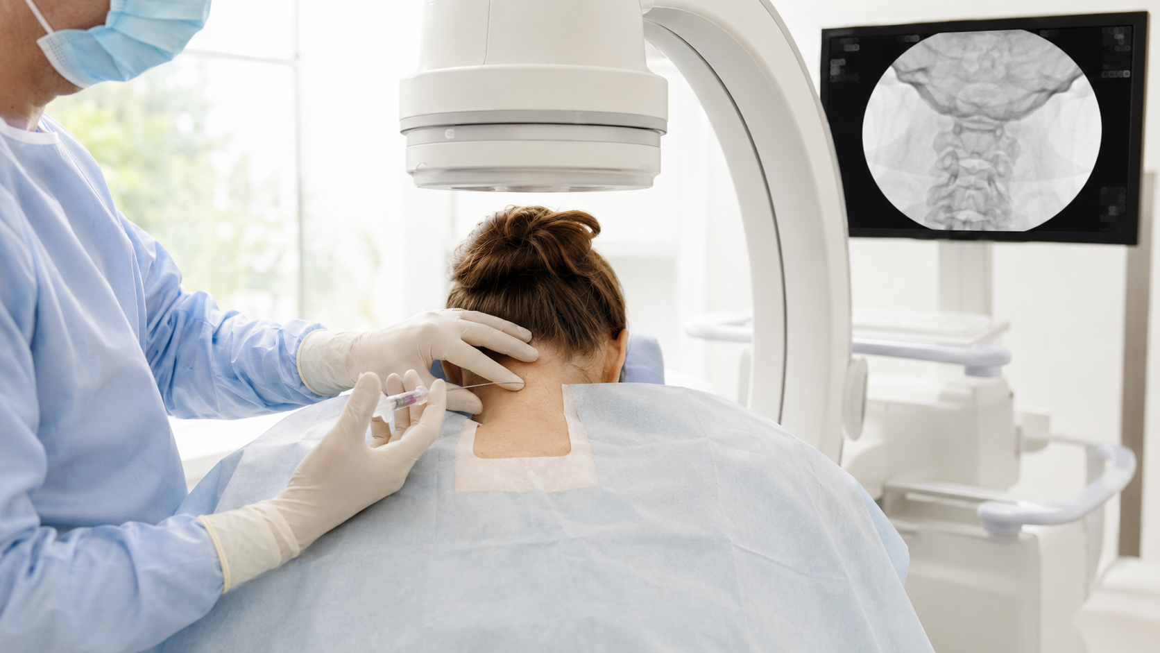
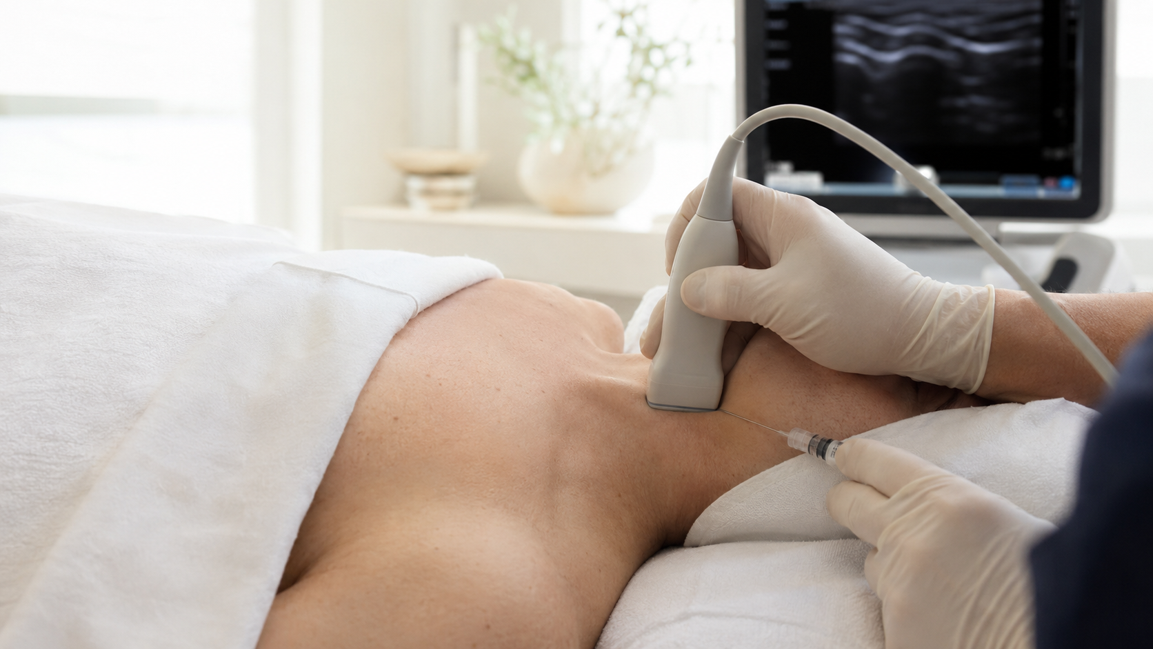
For example, trigger point injections may be considered when myofascial pain is a meaningful contributor. C1/2 facet injections may be considered only when the upper cervical facet joint is strongly suspected to be a pain generator. Ultrasound-guided injections may be used for selected peripheral, soft tissue, or musculoskeletal pain sources.
These procedures do not treat intracranial hypertension itself. They should not be presented as substitutes for neuro-ophthalmology, neurology, neurosurgery, imaging, lumbar puncture, CSF pressure evaluation, or urgent care when those are needed.
Stellate ganglion blocks, prolotherapy, PRP, BMAC, regenerative medicine, shunting, venous sinus stenting, optic nerve sheath fenestration, or other interventions should not be presented as universal treatments. Each requires diagnosis-specific evaluation, risk discussion, and specialist guidance.
When Specialty Care May Be Needed
Patients with suspected intracranial hypertension may need neuro-ophthalmology, neurology, neurosurgery, neuroradiology, ophthalmology, or primary care evaluation, especially when symptoms include papilledema, progressive visual symptoms, elevated opening pressure, abnormal imaging, venous sinus concerns, severe pressure headaches, or worsening neurologic symptoms.
MPM helps coordinate care when symptoms are complex and involve multiple possible contributors. This can be especially important for patients with EDS, HSD, POTS, MCAS-like symptoms, chronic migraine, CCI concerns, Chiari findings, CSF flow concerns, medical trauma, or long histories of unclear symptoms.
When Symptoms Require Urgent Evaluation
Patients should seek urgent evaluation for sudden severe headache, new weakness or numbness, vision loss, double vision, confusion, loss of coordination, fainting, trouble speaking or swallowing, new bowel or bladder dysfunction, fever, major trauma, severe progressive neurologic symptoms, chest pain, shortness of breath, or rapidly worsening symptoms.
Suspected papilledema or progressive visual change should be treated as urgent. These symptoms may reflect elevated intracranial pressure, stroke, infection, spinal cord compression, vascular emergency, traumatic injury, or another serious condition.
How MPM Approaches Intracranial Hypertension-Related Care
MPM approaches suspected intracranial hypertension through a diagnosis-first, coordinated model. The goal is to clarify the symptom pattern, review available imaging and eye findings, identify pain generators, avoid premature assumptions, and coordinate with the right specialists.
For patients looking for intracranial hypertension treatment in NYC, MPM offers a structured pain medicine perspective that considers pressure headache symptoms, chronic migraine, Chiari malformation, CSF outflow obstruction, CCI, upper cervical pain, EDS, HSD, POTS, MCAS-like symptoms, post-COVID pain, medical trauma, and complex chronic pain.
The goal is to help patients understand what may be contributing to their symptoms, identify appropriate next steps, and build a careful plan that supports safety, function, vision protection, and coordinated care.